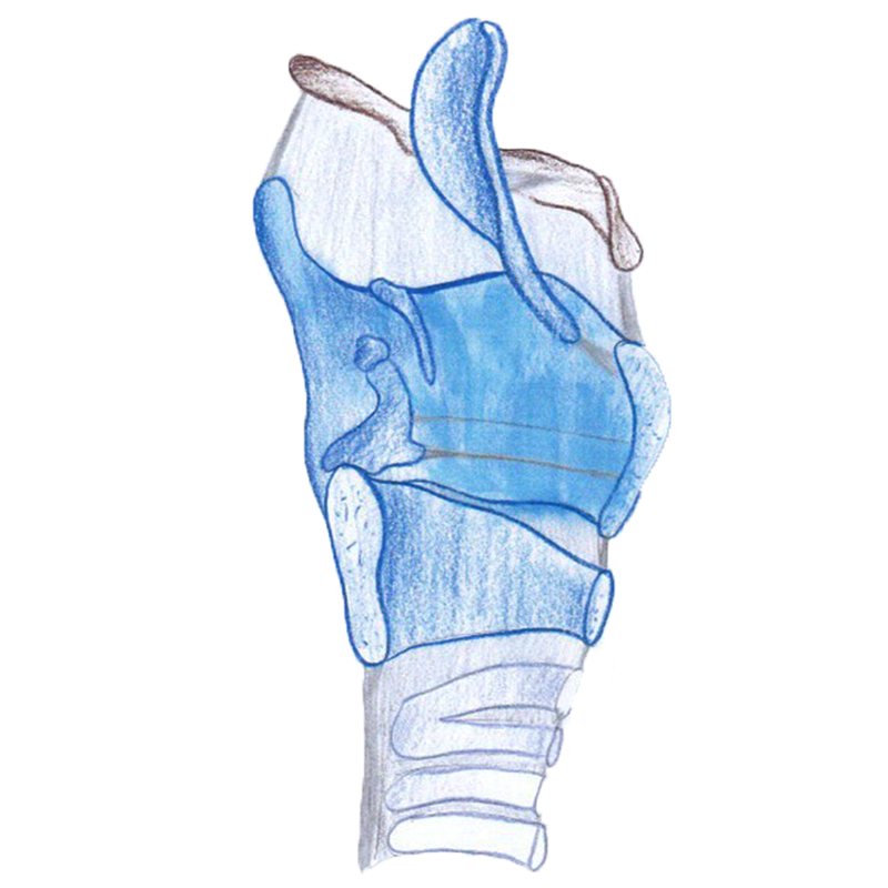
At the beginning of the transcutaneous LEMG examination, main structures of the anterior neck are palpated to identify the midline, the cricoid cartilage, the lower border of the thyroid cartilage, the thyroid notch, and the hyoid bone.
In obese patients or after neck surgery, palpating these landmarks can be difficult. With ultrasound, the landmarks can be localized and marked with a felt pen. If local anesthesia is used, excessive injections should be avoided to allow continued palpation of structures after injection.
If the patient has a tracheotomy, it is usually necessary to remove the tracheotomy tube to be able to place the needle. If the patient does not tolerate a short-term removal, a nasal speculum may dilate the tracheostomy and maintain the airway open during the examination.

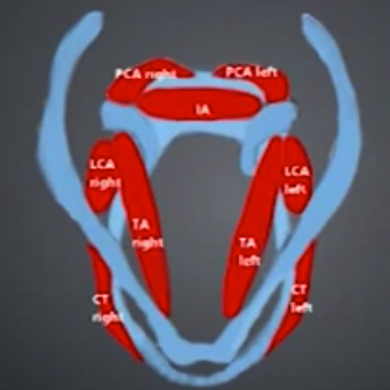
It is recommended to begin the LEMG with the
- thyroarytenoid muscle (TA), followed by an examination of
- the PCA, if there is a good level of tolerance.
Both muscles are innervated by the inferior recurrent laryngeal nerve (ILN). Finally, the needle should be inserted into the
- cricothyroid muscle (CT) to examine the function of the superior laryngeal nerve (SLN).
