The morphology comprises following characteristics of a motor unit action potential (MUAP):
MUAP Duration

The duration depends on the velocity of the neural input, which in turn is influenced by the insulation of the nerve. Nerves that are well insulated and have an intact and functioning sheath are able to transmit electrical impulses faster, because electrical impulses are transmitted from one node of Ranvier to another, the so called saltatory conduction.
The majority of reference EMG data are obtained from big skeletal muscles. However, this data cannot be directly compared with EMG signals of smaller muscles, particularly of the laryngeal muscles.
Latter show a shorter duration than bigger muscles, lasting ca. 5-6 ms, depending also on the needle used. The reason for a shorter duration and a smaller amplitude is the fact, that laryngeal muscles have motor units with a high innervation ratio. This means that every nerve fiber supplies only a smaller number of muscle fibers.
MUAP Amplitude

In contrast to the duration, the amplitude of the motor unit potential reflects the number and the strength of the muscle fibers innervated by one nerve ending.
A motor unit potential of a laryngeal muscle usually shows an amplitude of 200-500mV.
MUAP Shape
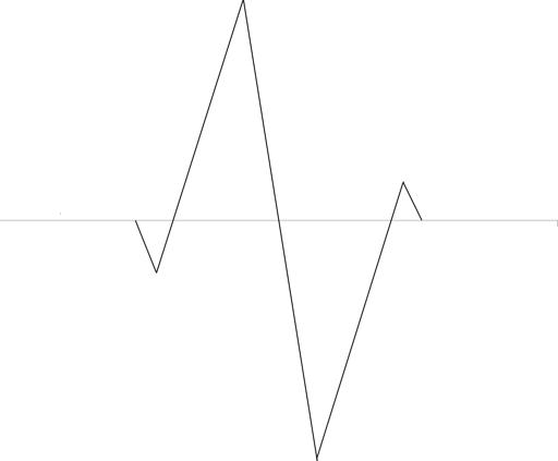
The shape of the motor unit potential reflects changes in the electrical activity of the muscle membrane.
Normally measured in a certain distance to the motor end plates, it has a biphasic pattern with an upward positive spike and a downward negative spike.
MUAPs recorded close to motor end plates appear often polyphasic. In the small laryngeal muscles this is hardly avoided. Nevertheless, the waveform morphology of the motor unit potential provides along with other information the phase of recovery. After injury, the nerve goes through a process of denervation, followed by regeneration:
- During denervation, there is no neural input into the muscle, and therefore no abnormal waveforms are produced.
- Abnormal motor unit potential morphologies are produced during the period of regeneration:

During the early phases of regeneration, tiny nerves return to the muscles, atrophied during the time of denervation. Early in the regrowth process, the insulation of the nerve is decreased. The combination of the tiny, minimally insulated nerves, and the weak muscle fibers produces electrical signals on the LEMG that are seen as motor unit potentials with small amplitudes, long durations, and polyphasic shapes. These waveforms are sometimes referred to as nascent units and imply the presence of a recent nerve injury.

During regeneration, the nerves become healthier and better insulated through regrowth of their sheaths. The muscle fibers become stronger and gain mass. However, not all nerve fibers regenerate. Fibers that do regenerate usually branch more extensively than they did before the injury, and they spread to innervate as many denervated muscle fibers as possible. The motor unit potentials that are produced as a result of this ongoing regeneration have greater amplitudes than normal. These motor unit potentials have a prolonged duration and are often described as giant polyphasic potentials. Previous nerve injury can be inferred from their presence.
- If the nerve is not injured but the muscle is damaged, the morphology of the motor unit potential is different. The intact nerve functions well, i.e., the duration of the motor unit potential is normal. Instead, the electrical charges in the muscle membrane are abnormal resulting in a polyphasic shape. The decreased amplitude reflects the decreased muscle mass and force of contraction.
As mentioned above, detecting these criteria in LEMG is complicated by the physiological background activity and the physiological appearance of polyphasic MUAPs caused by the small distance to the motor end plates.
The waveforms can be classified into:
- Normal biphasic motor unit potential
- Early (sometimes polyphasic) reinnervation potentials with low amplitude and long duration
- Giant polyphasic reinnervation potentials with high amplitude and long duration
- Myogenic polyphasic potentials with low amplitude but normal duration