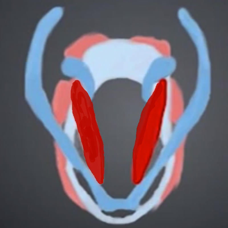
The cricothyroid notch, also known as cricothyroid space or cricothyroid membrane region, is the anatomic reference for the needle insertion.
First, the needle pierces the skin in midline in sagittal direction and will be placed directly under the lower border of the thyroid cartilage.
Second, the needle tip will be angled laterally (30°) and superiorly (15°) and penetrates through the cricothyroid ligament without entering the airway. It is also possible to place the needle tip around 5 mm laterally of the midline. In this case, the needle should be angled 20° laterally and 15° superiorly. Depending on the thickness of the neck and the entry angle, the thyroarytenoid muscle should be reached after pushing the needle 15 mm through the ligament.
Coughing of the patient generally indicates that the needle has penetrated the airway and is causing irritation of the mucosa. A burst of sine waves modulated by phonation also indicates that the electrode tip has entered the airway. In both cases, the needle should be withdrawn to the cricothyroid ligament and reinserted more laterally.
EMG Activity
- The position of the needle is confirmed by asking the patient to say /i:/ (‘ee’) or holding his breath by a glottal stop. During both procedures, EMG activity sharply and sustainably increases.
- Furthermore, swallowing causes a short strong thyroarytenoid activity during the glottal stop.
- While deeply breathing in and out, the resting activity drops periodically during expiration.
- If the needle is placed too laterally and records the activity of the lateral cricoarytenoid muscle (LCA), EMG activity increases and rapidly drops during phonation. Viewed from the cricothyroid space, the TA and LCA are nearly on the same axis and in direct contact to each other. It is thus not always possible to reliably differentiate between these two muscles. However, this is not necessary for most clinical questions.
Using a fiberoptic scope, moving the needle within the substance of the thyroarytenoid muscle and observing the vocal fold movement confirms the position of the electrode. This procedure may cause the patient to swallow or cough.
Click here to go back to Transcutaneous LEMG.